



Contents
Scroll to:
https://doi.org/10.25207/1608-6228-2026-33-2-15-26
Scroll to:
Background. Described more than half a century ago, the countercurrent multiplication mechanism in the loop of Henle remains hypothetical; it suggests that tubular flow is significantly influenced by physicochemical processes that have yet to be quantified. Objective: To develop a model of tubular fluid dynamics and establish a numerical criterion, where a decrease below unity (threshold value) indicates pathological flow deceleration (tubular stasis) in the loop of Henle, leading to subsequent metabolic stress on the epithelial cells.
Methods. The study used mathematical modeling to account for specific flow characteristics within nephron tubules; given the absence of smooth muscle, tubular fluid dynamics are driven primarily by osmotic (electrochemical) effects.
Results. The findings provide a means to quantify (numerically or computationally) the flow capacity of the loop of Henle. This issue has long attracted the attention of specialists, most of whom support the hypothetical countercurrent multiplication in the ascending limb. This mechanism assumes passive Na+ efflux from the tubular lumen to the interstitium to maintain the longitudinal osmotic gradient, which drives the tubular fluid dynamics.
Conclusion. The results suggest that various factors indirectly slowing the tubular fluid dynamics (such as lymphostasis and venous stasis) impede the passive Na+ efflux from the ascending limb. This triggers metabolic activation in the epithelial cells in this region, increasing the facultative production of adenosine triphosphate to drive active lumen-to-interstitium Na+ efflux. Chronic metabolic stress (catabolism) remodels mitochondrial biogenesis in the epithelial cells of the loop of Henle, creating conditions for the formation of calcium phosphate aggregates in the basal labyrinth.
Tatevosyan A.S., Bunyakin A.V., Alekseenko S.N., Katani Z.O., Yuldashev A.A., Ismatov B.A. A predictive model for the quantitative assessment of intratubular urodynamics during the creation of osmotic (electrochemical) gradient by the loops of Henle in the renal parenchyma. Kuban Scientific Medical Bulletin. 2026;33(2):15-26. https://doi.org/10.25207/1608-6228-2026-33-2-15-26
Renal function relies on the synergy between the glomeruli and the tubule system. While clinical assessments typically prioritize glomerular health, tubular processes are often underestimated, despite playing a fundamental role in everyday practice. Therefore, gaining a thorough understanding of the physiological mechanisms underlying tubular function is essential to grasping their relevance to common pathologies.
In urolithiasis patients, scanning electron microscopy of the kidneys reveals calcification within the basement membranes of the loop of Henle (LoH) in early-stage Randall’s plaques, extending into the adjacent papillary tissue [1]. While the exact mechanism of calcium phosphate (CaP) deposition in the basement membrane of thin LoH segments remains elusive, evidence suggests that Randall’s plaque formation originates in the ascending thin limbs [2]. It is hypothesized that calcium diffusion from the descending thin limb is the primary source of CaP supersaturation in the adjacent interstitium. In fact, the basement membranes of both the thin and thick LoH segments consist of collagen and mucopolysaccharides, forming an electrostatically charged matrix that readily attracts CaP, leading to progressive calcification.
The renal tubule system lacks a smooth muscle apparatus, suggesting that fluid dynamics are driven by physicochemical (electrochemical) processes. Although the countercurrent multiplication mechanism in the LoH was first described over half a century ago, it remains largely theoretical. The lack of a “numerical criterion” for LoH flow transport (https://pubmed.ncbi.nlm.nih.gov/?term=Numerical+criterion+of+flowing+transport+of+the+loop+of+Henle) further indicates that the underlying mechanism of tubular fluid movement has yet to be fully elucidated. A deeper understanding of the electrochemical processes within LoH epithelial cells, which drive fluid dynamics in the renal tubule system, will clarify the pathogenetic context of nephrolithiasis (nephrocalcinosis). This is supported by morphological evidence showing that initial CaP agglomerates originate in the interstitium near the LoH bend [3][4].
The study aims to develop a model of tubular fluid dynamics and to establish a numerical criterion, where a decrease below unity (threshold value) indicates pathological flow deceleration (tubular stasis) in the LoH, leading to subsequent metabolic stress on the epithelial cells.
The most common predictor of impaired (slowed) fluid dynamics in the renal tubule system is lymphostasis (venous stasis). In order to maintain these dynamics, the character of electrochemical interactions shifts, and energy metabolism is activated within LoH epithelial cells. For a quantitative (numerical) description of electrochemical interactions, a model was developed to derive a dimensionless diagnostic criterion. This criterion integrates quantities of diverse physical dimensions (temperature T in the renal parenchyma; flow rates Q0, Q1, and Q2 , across LoH segments; osmolality difference c1 – c2 = μΔc (see below for detailed explanations), which makes it more informative (effective) than any single parameter alone.
The study draws on a broad range of physiological and biochemical data regarding nephron function, the specific flow characteristics in the descending and ascending LoH limbs, and the anatomical features of the tubular endothelium.
Physiologists and morphologists participating in the study note that the initial stage of nephrolithiasis is characterized by basement membrane calcification in the LoH; however, no satisfactory explanation for this phenomenon exists. We propose a mathematical model of tubular fluid dynamics that explains the mechanism underlying metabolic stress on LoH epithelial cells. This stress disrupts mitochondrial biogenesis, contributing to the deposition of calcium phosphate (CaP) salts in the basal labyrinth of the thin LoH. This model will undoubtedly provide valuable insights for clinicians interested in the pathogenesis of nephrolithiasis (nephrocalcinosis) and may define the direction of future research.
The study aims to develop a model for a numerical criterion whose decrease below a threshold of unity indicates pathological flow deceleration (tubular stasis) in the LoH. Specifically, the objectives are to identify the primary effects involved in the passive transport of Na⁺ within the renal parenchyma that can be mathematically modeled with acceptable accuracy; to establish the core numerical characteristics and equations (physical laws) modeling these phenomena, i.e., to construct a mathematical LoH model that would be sufficiently simple to avoid cumbersome calculations yet adequate for diagnostic use; to derive (based on these equations) a dimensionless criterion and establish its approximate baseline value for the kidney’s normal function as an “osmotic pump” driven by Na⁺ transport within the renal parenchyma.
This study builds upon the long-standing hypothesis of a countercurrent multiplication mechanism in the LoH. While this hypothesis explains the general features of tubular fluid dynamics, it remains a qualitative framework. It fails to factor in the quantitative indicators of reduced LoH flow capacity, a condition that increases metabolic demands (adenosine triphosphate production) within epithelial cells.
The main research focus is the interrelationship between the variables comprising the proposed dimensionless criterion. However, it is challenging to estimate statistical sample sizes, as such data are often omitted in the studies upon which this meta-analysis and the present study are based.
Instead of statistical analysis, this study employed fundamentally different methods for mathematical modeling of fluid dynamics (and associated electrochemical effects) within the LoH. This key nephron component functions as a system for generating a pressure gradient based on osmotic principles rather than mechanical action (myocytes). The methodology is characterized by the following features: 1) the study focuses on nephron physiology, biochemical processes within the tubular endothelium, and the specific fluid dynamics in the descending and ascending LoH limbs; 2) the study period corresponds to the chronology of cited publications, as the data set was compiled through the meta-analysis and systematization of available information; 3) the evaluated parameters are integrated into a new (previously unpublished) numerical criterion, where a decrease below a threshold of unity indicates flow deceleration in the LoH; 4) direct measurement methods for estimating the percentage of functional LoH units within the renal parenchyma are virtually nonexistent. This underscores the relevance of the present study.
No risk groups were established in this study. However, it is recognized that the longitudinal osmotic pressure gradient is the primary driver of fluid movement (primary and secondary urine) within the LoH. To assess this process, it is essential to quantify the flow parameters and their interrelationships. This approach yields a dimensionless criterion that reflects the functional state of tubular fluid dynamics.
In recent decades, efforts have been made to deepen the understanding of the relationships between the renal tissue structure, function, and endophenotype in order to gain insight into kidney physiology and pathophysiology [5]. At the tissue level, the functional organization of kidneys involves distinct cell subtypes tailored to specific physiological needs [6]. With the application of Omics technologies, it has become possible to map the structural and functional characteristics of nephron epithelial cells across various segments of the tubular system.
The tubular fluid flow within the nephron, occurring despite the lack of smooth muscle tissue, has always attracted the attention of specialists, most of whom support the countercurrent multiplication mechanism in the LoH. Each human kidney contains 8–12 medullo-papillary complexes that have a parabolic shape. This geometry implies the presence of short, peripherally located nephrons and long, centrally located nephrons. It is argued that the parabolic shape of these complexes determines their function and serves as a critical factor in the predisposition to biomineral formation. [7].
Each medullo-papillary complex is divided into two zones: cortical (1) and medullary (2). The single-layered cortical epithelium of the proximal and distal nephron segments is microvillous (rough), while the medullary epithelium of the LoH is smooth (squamous). In turn, the endothelium of the LoH contains several cell types having different reabsorption mechanisms [8], meaning that the LoH bend has functionally distinct segments: the descending (1) and ascending limbs (2).
Initial morphological changes in cellular structures are driven by the delicate balance between metabolic demand and oxygen supply, which varies significantly across renal regions. The functional heterogeneity of various nephron segments is reflected in their distinct metabolic pathways [9]. Comparisons of oxygen availability revealed nephron segments with high susceptibility to hypoxia-induced kidney injury [10]. Specifically, pО2 levels in capillaries supplying the microvillous epithelium of the proximal and distal tubules are 45–50 mm Hg, whereas in those supplying the squamous epithelium of the LoH, pО2 drops to 35–40 mm Hg. This reflects the divergence in energy metabolism (dissimilation) within these structures [11] (Fig. 1). Notably, the relative temperature in the medullary zone is higher than that in the cortical zone.

Fig. 1. Oxygen tension and temperature gradients at the renal corticomedullary junction (А). The osmotic gradient (mOsm/kg) in the renal parenchyma is directed toward the apex of the renal papilla, along the loop of Henle (B). The values 1200, 900, and 600 correspond to the section of the loop of Henle with the maximum osmotic gradient
Note: The figures were created by the authors.
Рис. 1. Градиент напряжения кислорода и температуры в корково-медуллярной зоне почечной паренхимы (А). Осмотический (мосм/кг) градиент в почечной паренхиме направлен в сторону верхушки почечного сосочка, вдоль ПГ (B). Значения 1200, 900, 600 соответствуют участку ПГ с максимальным осмотическим градиентом
Примечание: рисунки выполнены авторами.
The urine concentration mechanism is a primary kidney function, characterized by the generation of an osmotic gradient that increases toward the medulla. This gradient arises from the accumulation of dissolved substances, specifically NaCl and urea, within the cells, interstitium, tubules, and medullary vessels [11]. The mechanisms for the independent regulation of water and sodium primarily operate in the renal medulla. Here, medullary nephron segments and the vasa recta are organized in complex, specific anatomical relationships that depend on their three-dimensional configuration [12].
Renal circulation is characterized by the branching of the efferent arteriole into the cortical (greater) and medullary (lesser) systems, forming a capillary network that envelops the tubule system. This rete arterial mirabile primarily surrounds the ascending LoH limb, while the descending limb is enveloped by the efferent venous network (Fig. 1). The peritubular capillaries are uniquely adapted for reabsorption; following glomerular filtration, the increased concentration of formed elements significantly raises the oncotic pressure (by 8–12 mm Hg), while blood flow velocity in this segment remains minimal.
This pressure gradient drives tubular flow (ensuring tubular fluid dynamics). Such a mechanism can be described as an “osmotic pump” powered by the fourfold osmolality gradient within the interstitial tissue directed toward the apex of the medullary papilla (Fig. 2).

Fig. 2. The descending limb segment of the loop of Henle (~12 μm in diameter, spanning nearly the entire 2-cm thickness of the renal parenchyma) is impermeable to salts and urea but highly permeable to water, creating an osmotic (concentration) gradient (А). Under physiological conditions, passive (adenosine triphosphate-independent) efflux of Na⁺ cations occurs through intercellular spaces in the ascending limb, creating an electrochemical (osmotic) gradient (B)
Note: The figures were created by the authors.
Рис. 2. Сегмент нисходящего колена петли Генле (диаметр порядка 12 мкм, пронизывающий почти всю толщину почечной паренхимы порядка 2 см) непроницаем для солей и мочевины, но легко проходим для воды, что создает осмотический (концентрационный) градиент (А). В физиологических условиях в восходящем отделе ПГ осуществляется пассивное (без затраты энергии АТФ) выведение катионов Na⁺ через межклеточные промежутки, что создает электрохимический (осмотический) градиент (B)
Примечание: рисунки выполнены авторами.
The resulting osmotic gradient enables passive water reabsorption from the descending LoH limb into the interstitial space without adenosine triphosphate (ATP) consumption by epithelial cells: molecules in amorphous association with water selectively and passively permeate the descending limb walls into the interstitium for their subsequent return (reabsorption) into the bloodstream. This hydraulically passive segment exhibits decelerating flow and decreasing luminal pressure; meanwhile, the rising osmolality difference facilitates the accumulation of energy, which is later transformed and used by the “osmotic pump” of the ascending LoH limb (Fig. 2).
Subsequently, tubular fluid dynamics are driven by the hydraulically active ascending LoH limb, where pressure losses are compensated through an electrochemical (osmotic) mechanism. This is achieved by the efflux of sodium cations (Na⁺) into the interstitium. The single layer of epithelial cells lining the tubule walls effectively functions as a biological membrane and, under physiological conditions, allows Na⁺ to pass through the intercellular spaces passively, without ATP consumption. This process generates a predominantly positive charge on the interstitial side of this ascending segment.
Negatively charged chloride ions (Cl⁻) remain within the tubular lumen; as they undergo Brownian motion and collide with the tubule wall (biological membrane), without the ability to permeate it, they generate not only a transmural osmotic pressure difference (calculated via the Van’t Hoff equation1 ΔP0 = iΔc · R0T [13]), but also a longitudinal osmotic gradient.
The tubular fluid acidification is mediated by the chloride-independent Na/H antiporter in the luminal membrane and a voltage-dependent bicarbonate efflux mechanism in the basolateral membrane. Approximately 40% of NaCl reabsorption is passive, and 60% is active [14]. The potential difference between the tubular lumen and the epithelial cell affects chloride absorption. Notably, in convoluted tubules, active transport is electroneutral, resulting in an equivalent transfer of sodium and chloride across the cells [15].
Compared to the proximal and distal nephron segments, the epithelial cells lining the LoH possess a relatively sparse population of mitochondria, suggesting that their function requires minimal ATP-dependent mechanisms. Since Na⁺ efflux in this region occurs passively, it is reasonable to hypothesize that LoH mitochondria primarily function in thermogenic mode (as elaborated in the conclusion). This process may facilitate enhanced electrolytic dissociation, thereby maximizing the osmotic gradient along the LoH.
Epithelial cells in the thick ascending LoH limb contain a relatively larger mitochondrial pool, indicating ATP-dependent active transport. In cases where passive efflux in the ascending limb is compromised (e.g., by lymphostasis), Cl⁻ ions shift to electroneutral cotransport with Na⁺ [16]. However, from a general biosynergistic perspective, active Na⁺ transport in the thick ascending LoH limb can be viewed as a compensatory energy boost to the “osmotic pump” in the ascending limb where osmotic pressure declines. The objective of subsequent modeling is to demonstrate that passive Na⁺ transport is sufficient to maintain normal tubular fluid dynamics, provided a specific dimensionless criterion exceeds unity.
The criterion for evaluating LoH flow capacity during Na⁺ reabsorption is defined as the ratio of the longitudinal pressure difference across the ascending LoH limb to the pressure drop predicted by the Hagen–Poiseuille equation2 for laminar flow within the tubular lumen. The resulting relationship is as follows (a detailed derivation is provided below):

This formula uses the following constants and semi-constants:
π ≈ 3.14… — mathematical constant pi;
R0 ≈ 8.31  — universal gas constant;
T — absolute temperature (in Kelvin, normal for the human body and averaged across all organs 273.16 + 36.5 = 309.66);
η — viscosity of the solution (primarily water, approximately 0.001  ).
).
μ — molar mass of the dissolved salt (primarily NaCl) of 0.05844  .
.
Physiological variables are as follows:
ρ0, ρ1 — solution densities at the inlet (0) and outlet (1) of the hydraulically passive LoH segment ( ); ρ1, ρ2 — solution densities at the inlet (1) and outlet (2) of the active segment. For estimation purposes, these are assumed to equal the density of water: ρ0 = ρ1 = ρ2 = 10³
  (main urine component);
c1, c2 — mass concentrations of Na⁺ and Cl⁻ ions at the inlet and outlet of the hydraulically active LoH segment.
The calculations include intermediate steps and numerical substitutions to ensure that the method is reproducible via manual calculation, without the need for specialized software.
Since osmolality3 increases sharply in the descending LoH limb, the osmolality difference across this segment can reach 0.9 Osm/kg (as shown in Fig. 4): specifically, 1200 – 300 = 900 mOsm/kg = 0.9 Osm/kg;
Given that the osmolality of a 1 molal NaCl solution (1 mol of NaCl with a molar mass of 0.05844 kg/mol dissolved in 1 kg of water) is 2 Osm/kg, the difference in mass concentrations of the NaCl solution at the inlet (1) and outlet (2) of the ascending LoH limb comes to
c1 – c2 = (0.9/2) × 0.05844 = 0.026298 kg(NaCl)/kg;
Q0, Q1, Q2 — volumetric flow rates ( ) at the inlet (0) and outlet (1) of the hydraulically passive (descending) LoH segment, and at the outlet (2) of the active (ascending) segment, respectively. For estimation purposes, these values are assumed to be equal:

Here, SQ = 2·10⁻³ m³ represents the average daily urine output, approximately two liters of secondary urine processed per МН = 10⁶ normally functioning LoHs (assumed value);
L1 = 0.02 m; L2 = 0.015 m — lengths of the descending and ascending limbs, respectively: approximately 2 cm for the passive (1) segment and 1.5 cm for the active (2) segment;
d = 1.2·10⁻⁵ m — loop diameter (12 μm); flow velocity  ; thus, the flow travels approximately 17 diameters per second, and diffusion can be neglected: the convective term in the advection-diffusion equation is significantly larger than the diffusive term:
 ,
,
that is,

where
 — the coefficient of NaCl diffusion in water;
L = 0.01 m — characteristic linear dimension along the LoH;
Δc = (c1 – c2) / μ — difference in molar osmotic concentrations ( ). As calculated above, the mass concentration difference is c1 – c2 = 0.026298 kg(NaCl)/kg; i — isotonic coefficient (or Van’t Hoff factor) is defined by the theoretical formula i = 1 + α(n – 1) > 1, where α is the degree of dissociation and n is the number of ions or atoms with polar bonds that dissociate in solution. In practice, the isotonic coefficient is always lower than its theoretical value. For instance, for a 0.05 molal NaCl solution, the actual value of i is 1.9, compared to a theoretical i = 2.0, while for a 0.05 molal magnesium sulfate solution, i is 1.3. For the following estimation, i is assumed to be 1.5; ΔP1 — pressure drop for Poiseuille flow along the descending LoH segment (length L1), with flow rates Q0 and Q1 at its inlet and outlet (0, L1):
 .
.
Taking the difference in density at the segment ends into account,
 .
.
Similarly, pressure drop in the ascending LoH segment (length L2), with the flow rates Q1 and Q2 at its ends, is as follows:
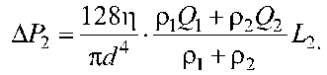
The pressure loss across the entire loop is expressed as:
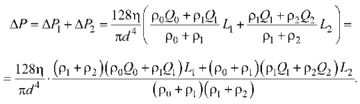
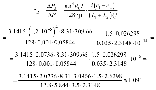
Based on the parameters listed above (items 1–5), an order-of-magnitude estimation of LoH flow capacity is as follows:
Thus, while the tubular walls are elastic, they lack smooth muscle tissue, rendering peristalsis a negligible factor in fluid dynamics. Peristaltic movement may only occur due to external stimuli. Consequently, the LoH primarily functions as an “osmotic pump.”
A specific feature of the model is that the numerical expression for the criterion τd (LoH flow capacity) given below uses a ratio of very small quantities, with the resulting value of τd being close to unity. The physical parameters used in the estimation are physiologically grounded rather than arbitrary, representing average values for normally functioning kidneys. To demonstrate this, we provide detailed calculations, including the substitution of input parameters (expressed in SI units).
The efficiency of this model is quite high, as calculating the criterion τd requires monitoring of only three parameters: the average temperature T within the renal parenchyma; the flow rates Q0, Q1, and Q2 in LoH segments (these can be considered proportional to fixed coefficients, meaning only daily urine output needs to be tracked); osmolality difference c1 – c2 = μΔc, which correlates well with the pH factor of secondary urine.
The limitations and assumptions of the model stem from the inevitable inaccuracy of input data parameters, specifically: average renal parenchymal temperature T, which requires 3D thermal imaging for accurate assessment; flow rates Q0, Q1, and Q2, whose proportionality coefficients may vary, potentially reducing the accuracy of estimations based solely on daily urine output; osmolality difference c1 – c2 = μΔc may change during long-term observation, while values tracked via correlation with the pH of secondary urine may involve statistical errors. Measurement of densities (such as ρ0, ρ1, and ρ2) is challenging in a clinical setting; assuming they equal the density of water introduces an inaccuracy, albeit a priori minor.
Since Na⁺ efflux in the ascending LoH limb can occur via paracellular pathways (predominantly, without ATP consumption), we hypothesize that in these epithelial cells, the mitochondrial electrochemical potential (ΔΨm) generated across the inner membrane is predominantly dissipated as heat production. This, in turn, facilitates the “osmotic pump” mechanism that drives tubular fluid dynamics.
This heat production mechanism is mediated by mitochondria during the first reverse cycle (F-I ⇋ F-IV), as conceptually described in [17][18]. Noteworthy is that an excessively prolonged (chronic) predominance of this cycle in mitochondrial biogenesis is accompanied not only by increased heat production, but also by the accumulation of amorphous CaP salts within the matrix. These salts can subsequently crystallize into CaP minerals in the basal labyrinth of LoH epithelial cells, acting as a triggering factor in the pathogenesis of nephrolithiasis (nephrocalcinosis).
The present authors conclude that the molecular profiles of metabolic pathways serve as a basis for understanding and predicting hypoxia-induced kidney injury in clinical settings. Overall, the prevalence of mitochondrial dysfunction in the renal population has been underestimated, as certain nephron regions exhibit increased susceptibility to mitochondrial dysfunction and subsequent toxin-induced damage [17][18].
Mitochondrial biogenesis, as a dynamic system shifting between fusion and fission, is a crucial aspect of renal physiology. However, the current evidence regarding these phenomena does not fully explain why mitochondria exhibit such remarkable efficiency. Unlike man-made technical devices, mitochondria appear to lack energy losses in the form of wasted heat or friction. As the functional activity of individual mitochondria, mitochondrial biogenesis serves as an example of a “heat engine” far more complex than any engineered device, characterized by a cycle with two reverse sub-cycles. Of note is that heat is generated not only through the intermediate accumulation of energy as ΔΨm across the inner mitochondrial membrane (for adenosine diphosphate to ATP conversion); heat production is also conceptually possible directly within the mitochondrial matrix during the first reverse cycle F-I ⇋ F-IV [17][18].
Although the kidneys are characterized by a high metabolic rate and, consequently, high ATP consumption, energy production rates and pathways vary among the different epithelial segments of the LoH. For instance, in the thin segments, Na⁺ reabsorption occurs passively under physiological conditions.
However, during flow deceleration (tubular stasis), an energy-consuming ATPase mechanism is activated to maintain the LoH osmotic gradient. This process involves the electroneutral cotransport of Na⁺and Cl⁻ ions. In other words, various factors that impair fluid dynamics (e.g., lymphostasis) hinder the passive efflux of Na⁺ from the ascending limb. This imposes a metabolic load on the local epithelial cells, triggering compensatory ATP production to drive active Na⁺ removal from the tubular lumen and sustain the necessary flow within the tubular system.
Increased metabolic demand in LoH epithelial cells triggers a functional transition of mitochondria to the first reverse cycle (F-I ⇋ F-IV). This shift promotes the formation of CaP agglomerates within the mitochondrial matrix, while chronic metabolic stress (catabolism) leads to the calcification of the LoH basement membrane, followed by the development of nephrocalcinosis and/or nephrolithiasis.
This study represents a theoretical synthesis yielding a diagnostic criterion for the numerical assessment of LoH flow capacity. According to this criterion, a value of  indicates normal renal fluid dynamics relative to average parameters of a healthy individual. The dimensionless criterion τd incorporates at least three parameters that may vary depending on the patient or in a single patient over time: the temperature T that may be higher or lower than the reference value used for the calculation; the flow rates Q0, Q1 and Q2; the osmolality difference c1 – c2 = μΔc. The densities (such as ρ0, ρ1, and ρ2) and deep-tissue temperature T can be measured using fairly accurate ultrasound (probing) devices and 3D thermal imagers; the value of c1 – c2 = μΔc can occasionally be determined via puncture and considered constant over a long period (monitored via the correlation with secondary urine pH).
A decrease in τd below unity may serve as a precursor to various disorders associated with nephron function. In essence, the diagnostic procedure focuses on determining the number of normally functioning LoHs. For instance, the flow rate value used in this estimation (item 3) is assumed as a baseline, considering that the number of LoHs (MH = 10⁶) represents a standard reference for an average healthy individual:

Even if the flow rates Q0, Q1 and Q2 across different LoH segments are not uniform (when estimated from the daily urine output), they remain inversely proportional to MH. An approximate estimation of the differences between these flow rates, resulting from density variations across LoH segments, is given by:
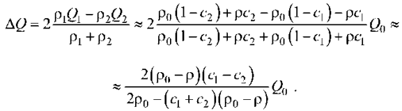
Consequently, the relative change in the volumetric flow rate is as follows:
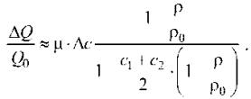
Given the above, c1 – c2 = μΔc = (0.9/2) · 0.05844 = 0.026298 kg(NaCl)/kg, and the relative density of NaCl to water  = 2.165, as well as assuming the limiting case of near-complete osmotic gradient dissipation c1 – c2 ≈ c1 + c2, we obtain an estimate of
  = –0.03.
Accordingly, the estimated value of the criterion is given by:

The sensitivity of this value to variations in the key input parameters is approximately one percent.
Establishing the functional reserve of the parameter τd is equivalent to determining the number of normally functioning LoHs. Since τd and MH are directly proportional, any deviation of MH from the baseline of 10⁶ results in a proportional deviation of τd from unity.
Following the concept of thermodynamic and electrochemical cyclicity in mitochondrial function [17–19], it is reasonable to hypothesize that heat production is primarily driven by the predominant operation of mitochondria in the first reverse cycle (F-I ⇋ F-IV), which is exergonic in nature. Conversely, in the cortical part of renal parenchyma, mitochondria operate mainly in the second reverse cycle (F-II ⇋ F-III). This cycle is endergonic, facilitating optimal ATP synthesis, while potentially leading to a localized cooling effect.
Although the details of this spatial organization of mitochondrial life cycles (biogenesis) have yet to be ascertained, the following calculations provide conceptual confirmation of this phenomenon.
According to the pressure drop formula for Poiseuille flow in the LoH:
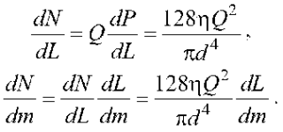
Here,  is the reciprocal of
  , the average number of mitochondria in the proximal tubule walls, normalized to one longitudinal meter (approximately 100 per cross-section);
  is the ATP power output of a single mitochondrion (W), which is used to drive fluid movement in the LoH (in its ascending segment; for the entire loop, the estimated value of
  can be doubled). Based on the parameters specified above (items 1–5), we obtain:

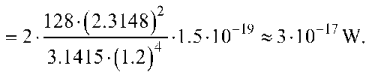
This value is approximately 1,000 times smaller than the average ATP energy output of a single mitochondrion [18][19].
Drawing on the Hagen—Poiseuille equation, modulation of tubular and vascular fluid viscosity (urine/blood) could enhance tubular fluid dynamics, thus mitigating the risk of amorphous CaP aggregate deposition.
The obtained result represents a dimensionless composite criterion, whose deviation below unity serves as a diagnostic indicator of impaired renal fluid dynamics. In the event of tubular stasis, energy metabolism is activated in this region to maintain the longitudinal osmotic gradient that drives tubular flow, while chronic metabolic stress (catabolism) in LoH epithelial cells remodels mitochondrial biogenesis, creating conditions for CaP agglomerate formation.
The significance of these findings lies in the fact that mathematical modeling provides a quantitative assessment of the normal tubular flow function. The “osmotic pump” mechanism, driven by Na⁺ transport within the renal parenchyma, can now be characterized by a single diagnostic criterion: τd >1. In addition to its practical applications, this result raises some profound theoretical issues, namely: compared to the proximal and distal tubular segments, the inner surface of the LoH is lined with squamous epithelial cells of minimal size and low mitochondrial content, indicating a significant reduction in energy-intensive secretory and reabsorptive processes in the LoH. In order to determine the pathways of energy production in various renal epithelial cells, a targeted metabolic ontology was developed, which enabled the differentiation between their catabolic and anabolic nature [19–21].
The practical application of this study can be described as prognostic, with results that can be used for diagnostic purposes in pathologies leading to impaired tubular flow dynamics, loss of renal function, and the development of nephrolithiasis (nephrocalcinosis) [22–24].
A deviation from the criterion τd >1 reliably reflects an activation of energy metabolism within the epithelial cells of the ascending LoH limb. This compensatory response serves to maintain the longitudinal osmotic gradient driving tubular flow; however, chronic metabolic stress (catabolism) induces a remodeling of mitochondrial biogenesis in these cells, creating a favorable environment for CaP aggregate formation in the basal labyrinth.
1. Yershov Yu. A., Popkov V. A., Berlyand A. S. General Chemistry. Biophysical Chemistry. Chemistry of Biogenic Elements. Moscow: Vysshaya Shkola, 1993 (Reprint 2023 ISBN 978-5-9916-8660-0978-5-9916-8661-7), see pp. 540–541.
2. Ebert G. A concise handbook of physics: A reference edition. Edited by K.P. Yakovlev. Translated from the 2nd German edition by N.M. Shikunina. Moscow: Fizmatgiz, 1963.
Akhmetov N.S. General and inorganic chemistry. Section III, States of Matter. Solutions. 2001. ISBN 5-06-003363-5 (Vysshaya Shkola), ISBN 5-7695-07-04-7 (Akademkniga).
3. Ibid.
1. Mager R, Neisius A. Aktuelle Konzepte zur Pathogenese von Harnsteinen [Current concepts on the pathogenesis of urinary stones]. Urologe A. 2019;58(11):1272–1280. German. https://doi.org/10.1007/s00120-019-1017-z
2. Evan AP, Coe FL, Lingeman J, Bledsoe S, Worcester EM. Randall’s plaque in stone formers originates in ascending thin limbs. Am J Physiol Renal Physiol. 2018;315(5):F1236–F1242. https://doi.org/10.1152/ajprenal.00035.2018
3. Bushinsky DA. Nephrolithiasis: site of the initial solid phase. J Clin Invest. 2003;111(5):602–605. https://doi.org/10.1172/JCI18016
4. Evan AP, Worcester EM, Coe FL, Williams J Jr, Lingeman JE. Mechanisms of human kidney stone formation. Urolithiasis. 2015;43 Suppl 1(0 1):19–32. https://doi.org/10.1007/s00240-014-0701-0
5. Dantzler WH, Layton AT, Layton HE, Pannabecker TL. Urine-concentrating mechanism in the inner medulla: function of the thin limbs of the loops of Henle. Clin J Am Soc Nephrol. 2014;9(10):1781–1789. https://doi.org/10.2215/CJN.08750812
6. Pannabecker TL, Dantzler WH, Layton HE, Layton AT. Role of three-dimensional architecture in the urine concentrating mechanism of the rat renal inner medulla. Am J Physiol Renal Physiol. 2008;295(5):F1271–1285. https://doi.org/10.1152/ajprenal.90252.2008
7. Alamilla-Sanchez M, Alcalá Salgado MA, Ulloa Galván VM, Yanez Salguero V, Yamá Estrella MB, Morales López EF, Ramos García NA, Carbajal Zárate MO, Salazar Hurtado JD, Delgado Pineda DA, López González L, Flores Garnica JM. Understanding Renal Tubular Function: Key Mechanisms, Clinical Relevance, and Comprehensive Urine Assessment. Pathophysiology. 2025;32(3):33. https://doi.org/10.3390/pathophysiology32030033
8. Park J, Liu CL, Kim J, Susztak K. Understanding the kidney one cell at a time. Kidney Int. 2019;96(4):862–870. https://doi.org/10.1016/j.kint.2019.03.035
9. Hansen J, Sealfon R, Menon R, Eadon MT, Lake BB, Steck B, Anjani K, Parikh S, Sigdel TK, Zhang G, Velickovic D, Barwinska D, Alexandrov T, Dobi D, Rashmi P, Otto EA, Rivera M, Rose MP, Anderton CR, Shapiro JP, Pamreddy A, Winfree S, Xiong Y, He Y, de Boer IH, Hodgin JB, Barisoni L, Naik AS, Sharma K, Sarwal MM, Zhang K, Himmelfarb J, Rovin B, El-Achkar TM, Laszik Z, He JC, Dagher PC, Valerius MT, Jain S, Satlin LM, Troyanskaya OG, Kretzler M, Iyengar R, Azeloglu EU; Kidney Precision Medicine Project. A reference tissue atlas for the human kidney. Sci Adv. 2022;8(23):eabn4965. https://doi.org/10.1126/sciadv.abn4965
10. Scholz H, Boivin FJ, Schmidt-Ott KM, Bachmann S, Eckardt KU, Scholl UI, Persson PB. Kidney physiology and susceptibility to acute kidney injury: implications for renoprotection. Nat Rev Nephrol. 2021;17(5):335–349. https://doi.org/10.1038/s41581-021-00394-7
11. Eveloff J, Bayerdörffer E, Silva P, Kinne R. Sodium-chloride transport in the thick ascending limb of Henle’s loop. Oxygen consumption studies in isolated cells. Pflugers Arch. 1981;389(3):263–270. https://doi.org/10.1007/BF00584788
12. Eveloff J, Kinne R. Sodium-chloride transport in the medullary thick ascending limb of Henle’s loop: evidence for a sodium-chloride cotransport system in plasma membrane vesicles. J Membr Biol. 1983;72(3):173–181. https://doi.org/10.1007/BF01870584
13. Subramanya AR, Ellison DH. Distal convoluted tubule. Clin J Am Soc Nephrol. 2014;9(12):2147–2163. https://doi.org/10.2215/CJN.05920613
14. Gadzhiev NK, Gelig VA, Kutina AV, Gorgotsky IA, Karpishchenko AI, Gorelov DS, Semenyakin IV, Zakutsky AN, Kuleshov OV, Shkarupa DD. Urinary pH: its regulation and relevance in urolithiasis metaphylaxis. Urology Herald. 2022;10(4):120–140 (In Russ.). https://doi.org/10.21886/2308-6424-2022-10-4-120-140
15. Levitskaya ES, Batiushin MM. Kidney Tubules — Scientific and Applied Value. The Russian Archives of Internal Medicine. 2022;12(6):405–421. (In Russ.). https://doi.org/.20514/2226-6704-2022-12-6-405-421
16. Rector FC Jr. Sodium, bicarbonate, and chloride absorption by the proximal tubule. Am J Physiol. 1983;244(5):F461–471. https://doi.org/10.1152/ajprenal.1983.244.5.F461
17. Tatevosyan AS, Alekseenko SN, Bunyakin AV. Thermodynamic and electrochemical oscillations in the mitochondrial life cycle (biogenesis) — predictors of tissue calcification. Russian Journal of Physical Chemistry. 2024;98(1):159–168. https://doi.org/10.31857/S0044453724010203
18. Tatevosyan AS, Bunyakin AV, Alekseenko SN, Katani ZO. Thermodynamic and Electrochemical Characteristics of Urine Protein Molecules That Affect the Formation of Stones. Biochemistry (Moscow), Supplement Series B: Biomedical Chemistry. 2025;19(1):68–79. http://dx.doi.org/10.1134/s1990750824601346
19. Layton AT, Laghmani K, Vallon V, Edwards A. Solute transport and oxygen consumption along the nephrons: effects of Na+ transport inhibitors. Am J Physiol Renal Physiol. 2016;311(6):F1217–F1229. https://doi.org/10.1152/ajprenal.00294.2016
20. Hall AM, Unwin RJ, Hanna MG, Duchen MR. Renal function and mitochondrial cytopathy (MC): more questions than answers? QJM. 2008;101(10):755–766. https://doi.org/10.1093/qjmed/hcn060
21. Liu Y, Chen Y, Liao B, Luo D, Wang K, Li H, Zeng G. Epidemiology of urolithiasis in Asia. Asian J Urol. 2018 Oct;5(4):205–214. https://doi.org/10.1016/j.ajur.2018.08.007
22. Zverev YaF, Zharikov AYu, Brukhanov VM, Lampatov VV. Modulators of oxalate nephrolithiasis. inhibitors’ crystallization. Nephrology (Saint-Petersburg). 2010;14(1):29–49 (In Russ.). https://doi.org/10.24884/1561-6274-2010-14-1-29-49
23. Liu Y, Song L, Zheng N, Shi J, Wu H, Yang X, Xue N, Chen X, Li Y, Sun C, Chen C, Tang L, Ni X, Wang Y, Shi Y, Guo J, Wang G, Zhang Z, Qin J. A urinary proteomic landscape of COVID-19 progression identifies signaling pathways and therapeutic options. Sci China Life Sci. 2022;65(9):1866–1880. https://doi.org/10.1007/s11427-021-2070-y
24. Berger GK, Eisenhauer J, Vallejos A, Hoffmann B, Wesson JA. Exploring mechanisms of protein influence on calcium oxalate kidney stone formation. Urolithiasis. 2021;49(4):281–290. https://doi.org/10.1007/s00240-021-01247-5
Artur S. Tatevosyan — Dr. Sci. (Med.), Professor at the Department of Preventive Medicine and New Health-Saving Technologies, Professor at the Department of Urology.
Mitrofana Sedina str., 4, Krasnodar, 350063
Alexey V. Bunyakin — Cand. Sci. (Phys.-Math.), Assoc. Prof., Department of Oil and Gas Engineering and Land Management.
Svyazi str., 11, Yablonovsky, 385140
Sergey N. Alekseenko — Dr. Sci. (Med.), Prof., Head of the Department of Disease Prevention, Healthy Lifestyle and Epidemiology.
Mitrofana Sedina str., 4, Krasnodar, 350063
Zorik O. Katani — graduate student, Department of Urology.
Mitrofana Sedina str., 4, Krasnodar, 350063
Aziz A. Yuldashev — Director of the Fergana Branch.
Kadriyat str., 79, Fergana, 150102
Beksultan A. Ismatov — gradient student, Department of Urology and Andrology.
Furmanova str., 23, Bishkek, 72004
Tatevosyan A.S., Bunyakin A.V., Alekseenko S.N., Katani Z.O., Yuldashev A.A., Ismatov B.A. A predictive model for the quantitative assessment of intratubular urodynamics during the creation of osmotic (electrochemical) gradient by the loops of Henle in the renal parenchyma. Kuban Scientific Medical Bulletin. 2026;33(2):15-26. https://doi.org/10.25207/1608-6228-2026-33-2-15-26
 Email this article (Login required)
Email the author (Login required)
Email this article (Login required)
Email the author (Login required) Sedin str. 4, Krasnodar, Russian Federation, 350063
Tel.: (861) 268 55 02
E-mail: kubmedvestnik@ksma.ru, kubmedvestnik@mail.ru





